Wireless surface EMG is a field-deployable sport analysis tool, capable of capturing the neuromuscular data that no camera or force plate can provide.
Sprint mechanics are well-documented. Ground reaction forces, stride frequency, contact time and propulsive impulse have been measured, modelled, and linked to performance outcomes across decades of research. What remains systematically absent from most sprint assessments is the neuromuscular layer: how individual athletes recruit and coordinate their muscles to produce the kinematics that these models describe. A peer-reviewed study published in Sensors (Yokota & Tamaki, 2025) addresses that gap directly, using wireless surface electromyography (sEMG) to map agonist–antagonist muscle coordination across a full 50-metre sprint in ten trained athletes.
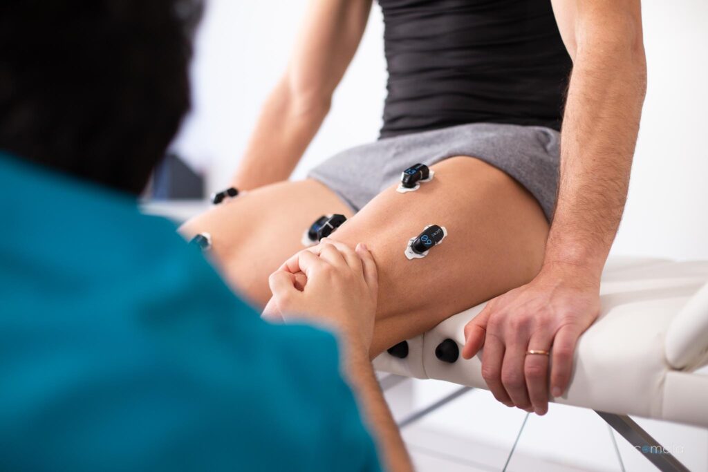
The instrumentation used was the Cometa Pico EMG wireless sensor system. The data it produced revealed something that no kinematic analysis could have retrieved: athletes performing the same task at comparable speeds were doing so with fundamentally different neuromuscular strategies. That distinction carries direct implications for performance optimisation and injury risk management in sprint athletes.
Why EMG Sport Analysis Covers the Blind Spot That Video Leaves Open
Video analysis and force plate data describe movement outcomes. They do not describe the neural strategy behind those outcomes. Two athletes can produce identical stride parameters while one relies on reciprocal inhibition between agonist and antagonist muscle groups and the other resolves the same mechanical demand through coactivation. The observable movement is indistinguishable. The neuromuscular cost and the ceiling for performance improvement differ substantially between the two strategies.
This is the practical problem that wireless sEMG solves in sprint analysis. Muscle activation timing and the relative contribution of antagonist pairs are not accessible through any other field-deployable measurement method. The study by Yokota and Tamaki establishes the methodological basis for making this data collection routine in applied sprint environments.
Study Design: 50-metre Field Sprint with Simultaneous EMG and Force Data
Ten trained sprinters (nine males, one female; mean age 21.8 years; mean maximal running speed 9.52 m/s) specialising in the 100 m, 200 m, 110 m hurdles, and 400 m hurdles performed four maximal 50-metre sprints from starting blocks on an indoor tartan track. The facility was equipped with 54 embedded force plates along the full sprint length, enabling simultaneous ground reaction force (GRF) acquisition at 2000 Hz, time-synchronised to the EMG signal via TTL trigger. This type of EMG and motion capture integration is increasingly viable in applied sport environments.
Surface EMG was recorded from four muscles of the right lower limb: biceps femoris (BF) and rectus femoris (RF) at the thigh, soleus (Sol) and tibialis anterior (TA) at the shank. These pairs represent the primary agonist–antagonist couplings involved in sprint mechanics at each joint level. Electrode placement followed SENIAM guidelines throughout. EMG signals were bandpass filtered at 10–500 Hz and sampled at 2000 Hz. All data were processed stride-by-stride in MATLAB, with integrated EMG (iEMG, expressed in %MVC·s) computed for each stride cycle across the full sprint distance.
Biceps Femoris in the Late Swing Phase: Performance Contribution and Injury Exposure
The most operationally relevant finding is the relationship between biceps femoris iEMG during the late swing phase and sprint velocity. Across all stride cycles, BF activation in this phase showed a significant positive linear correlation with running speed (r = 0.98, p < 0.05). The late swing phase corresponds to the backward motion of the leg just before foot strike, often referred to as pawing. Higher BF recruitment during this phase was associated with higher running velocity across all ten athletes.
BF activation in late swing did not show a significant correlation with mean anteroposterior force (mAP). The BF contribution in this phase therefore operates through swing-phase mechanics: faster backward limb motion and more rapid repositioning for the subsequent ground contact, rather than direct propulsive force at foot strike. This is consistent with the established understanding that hamstring function in sprinting is primarily eccentric and repositioning-oriented during late swing.
The injury implication follows directly. The late swing phase is where peak hamstring musculotendinous strain occurs during maximal sprinting, and where the majority of sprint-related hamstring injuries are initiated. An athlete producing higher BF activation to support better sprint mechanics is simultaneously operating under greater eccentric demand on the hamstring complex. Quantifying that exposure requires the EMG signal. It is not recoverable from kinematic or force data. The same principle applies across high-demand physical work contexts: see the role of EMG in physical risk assessment
Agonist–Antagonist Coordination: Consistent at the Ankle, Highly Variable at the Thigh
Cross-correlation function (CCF) analysis of the BF/RF and Sol/TA pairs across all stride cycles produced a structural contrast between the two joint levels that has direct implications for how sprint neuromechanics should be assessed and coached.
At the shank, Sol/TA coactivation was consistent across all ten participants. The mean CCF r-value at lag zero was 0.826 (SD 0.095), with a range of 0.395 to 0.980. The mean lag time between the two muscles was approximately zero (mean -0.019 s, SD 0.037), indicating near-simultaneous activation. This pattern reflects a shared strategy of ankle stiffening during the late swing phase in preparation for ground contact. It appeared consistently regardless of event specialisation or individual sprint mechanics, suggesting it represents a fundamental mechanical requirement rather than an individually optimised strategy.
At the thigh, the pattern was structurally different. The mean CCF r-value at lag zero for BF/RF was 0.645 (SD 0.124), with a range of 0.262 to 0.928. The mean lag time was 0.098 s (SD 0.008), with RF activation following BF. Phase-plane plots of individual BF/RF iEMG trajectories showed the full spectrum: some athletes produced clearly reciprocal activation profiles consistent with efficient alternation of hip and knee torques, while others showed markedly higher coactivation, consistent with a stability-oriented neural strategy under the extreme angular velocities of maximal sprinting.
These are not minor individual variations in the expression of the same underlying pattern. Athletes competing in the same events and producing comparable sprint velocities were operating with BF/RF coordination profiles distributed across nearly the full theoretical range. A coach observing these athletes from the trackside would have no basis for distinguishing their neuromuscular strategies from kinematic observation alone.
From EMG Sport Data to Training Decisions
The practical value of this data lies in the specificity it introduces into performance and injury management decisions. An athlete with high BF iEMG in the late swing phase and a strongly reciprocal BF/RF profile represents a different training case from an athlete with comparable BF activation and high thigh coactivation. Where reciprocal coordination is already established, the neuromuscular system is operating efficiently at high hamstring demand. Where coactivation predominates, unnecessary antagonist activity may be limiting propulsion output while simultaneously loading the hamstring complex. The sprint looks identical from the outside. The appropriate intervention requires the EMG data to distinguish between these two cases.
The authors note that the sample size of ten athletes limits statistical detection to strong correlations (|r| greater than or equal to 0.76), and that longitudinal work is needed to determine whether these coordination profiles are stable traits or adaptable through training. Both remain open questions. The methodological contribution is already established: wireless sEMG deployed across a full-length sprint track and synchronised with embedded force plate data produces the temporal and signal resolution required to characterise these differences stride-by-stride under field conditions.
Instrumentation: Pico EMG, Wave Plus, and EMG and Motion Tools
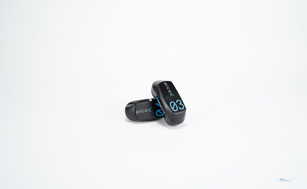
The sEMG data were acquired using Cometa Pico EMG wireless sensors, weighing approximately 7 g per unit, with integrated signal processing and wireless transmission to the Wave Plus receiver. Bipolar Ag/AgCl electrodes (8 mm diameter, 20 mm interelectrode distance) were applied following standard skin preparation to maintain impedance below 5 kOhm. Signals were bandpass filtered at 10–500 Hz and sampled at 2000 Hz, consistent with the Nyquist requirements for high-frequency sEMG content during ballistic contractions.
Data acquisition used EMG and Motion Tools (version 8.6.2.0), with synchronisation to the GRF system via TTL trigger generated by the starting pistol. Within-rater reliability for EMG amplitude at MVC was good across repeated trials, with an intraclass correlation coefficient of ICC(3,1) = 0.964.
The Information That Kinematics Cannot Provide
Sprint performance analysis has accumulated rigorous models of the mechanical and kinematic parameters that determine running speed. The neuromuscular layer that produces those parameters has remained largely inaccessible in field conditions. Yokota and Tamaki (2025) demonstrate that this layer is now measurable in applied environments, with sufficient temporal and signal resolution to characterise individual coordination strategies across a full competitive sprint distance.
The inter-individual variability in BF/RF coordination documented in this study is not measurement artefact. It is physiologically meaningful information about neural strategy and individual adaptation that kinematic analysis cannot retrieve. Two athletes produced comparable sprint velocities through substantially different neuromuscular strategies. Identifying which strategy each athlete is using, and whether it is the most efficient and sustainable one for that individual, is now a measurable question.
Ready to see the neuromuscular data behind your athletes’ mechanics?
Contact a Cometa specialist to discuss how wireless sEMG integrates into your existing performance assessment workflow.