In biomechanics and clinical gait analysis, we have spent decades perfecting the way we measure motion in human body and are able to perform many kinds of analysis through modern software, like our EMG & Motion tools. Optoelectronic, motion capture systems, and force plates, have allowed us to model joint kinematics and ground reaction forces precisely. These tools, however, describe the consequences of movement. Two patients can exhibit identical walking speeds and joint angles, but one might be operating under extreme antagonist coactivation while the other uses highly efficient reciprocal inhibition.
There is a blind spot: the neuromuscular layer, and to look into it researchers have long relied on surface electromyography (sEMG). We have already covered the use of sEMG in Gait Analysis, however, Today, as clinical and research paradigms shift toward personalised medicine, gait analysts face a technological transition: from Single Differential sEMG to High Density sEMG. Rather than viewing these technologies as competing, the most advanced gait laboratories are beginning to realise that using SD sEMG and High Density EMG in tandem provides a better picture of human locomotion.
Conventional Bipolar sEMG: The Clinical Workhorse of Temporal Coordination
Conventional bipolar sEMG is the clinical gold standard for in-field movement analysis. By measuring the difference in voltage between two electrodes placed over a muscle, it yields a clear, continuous, one-dimensional trace of global muscle activity.
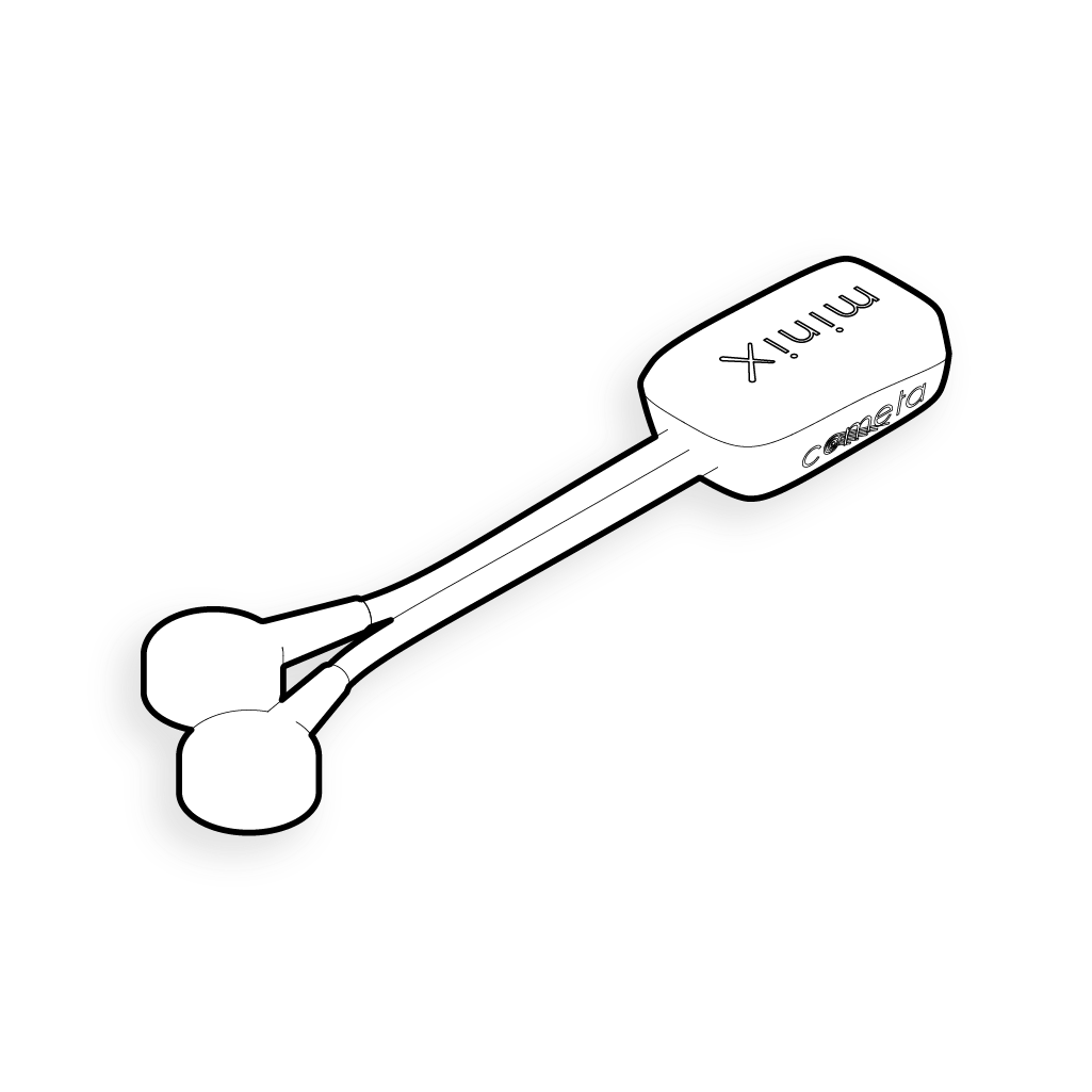
Strengths:
- electrodes are easier to place
- computationally lightweight
- Allows to record multiple muscle groups simultaneously across a full gait trial.
- It excels at identifying global muscle activation timing (on/off patterns) and mapping agonist-antagonist coordination profiles.
Limitations:
- large electrodes and higher interelectrode distances can act as a spatial low-pass filter, averaging out the high-frequency physiological details of the signal.
- Bipolar sEMG is highly susceptible to crosstalk (detecting signals from neighboring muscles, which is a major confound in small or pediatric muscle groups), and geometric artifacts.
- When walking, the muscle’s Innervation Zone (IZ) can shift directly beneath a fixed pair of bipolar electrodes, causing changes in signal amplitude that may be misinterpreted as changes in neural drive.
High Density sEMG: Mapping the Spatiotemporal Neural Drive
High Density EMG expands our view of muscle activity from a single temporal trace to a full spatiotemporal map. By employing grids of miniaturised electrodes (between 3 and 5 millimetres in diameter) with a tight interelectrode distance (IED between 5 and 8 mm) to prevent spatial aliasing, HD sEMG captures how electrical signals distribute across the skin over time, effectively creating a high-resolution animation of the activation of the muscle.
Strengths:
- wide spatial resolution
- facilitates tracking of muscle fibre conduction velocity (CV)
- Allows to precisely localise Innervation Zones, by applying spatial filters (like the Laplacian/NDD) it is possible to identify and remove crosstalk from adjacent muscles.
- Enables Motor Unit Decomposition (MUD): through advanced software algorithms it is possible to separate global muscle signals into single motor unit firing patterns.
Limitations:
- computationally intensive
- limited to isometric or highly controlled contractions: current BSS decomposition algorithms assume a stationary muscle state
How to implement both in gait analysis
The future of gait analysis goes towards a multi-channel approach, combining data from different tools: mocap, force plates and sEMG. Combining SD and HD sEMG provides one extra layer to this branch of biomechanics:
- The Macro-Level (Bipolar sEMG): During gait trials, wireless bipolar sensors (like our WaveX System) can be deployed across lower-limb muscle groups providing the overarching temporal scaffold of the walk. This will map joint kinematics and show muscular coactivation timing, and multi-muscle synergies across multiple stride cycles.
- The Micro-Level (HD sEMG): high density emg grids can be placed over specific target muscles of interest during controlled phases of the evaluation to dig deeper and find out how the central nervous system is controlling that activation.
By combining these layers, clinicians and researchers can measure the direct neural changes underlying gait rehabilitation. So for example, you can tell if an enhanced pattern of walking produced after some PT intervention reflects only a peripheral adjustment in joint angles, or if your patient was actually able to re-pattern their spinal motor neuron discharge frequencies and common input drives.